Radiology and Medical Imaging Sector
Introduction
The API /virtualbot/analisys_image_report/ is designed to support doctors and healthcare professionals in the analysis of medical images. This API processes sets of images related to a patient, combining them with their clinical history (if provided), and offers a preliminary diagnosis based on these images. The result is a detailed report of the findings in each image, along with personalized recommendations. It is important to note that the API does not store images or clinical history; it only retains the generated report, ensuring the privacy and confidentiality of patient information.
Functioning of the API /virtualbot/analisys_image_report/
Endpoint: POST /virtualbot/analisys_image_report/
Input Parameters:
- Patient Images: A .zip file containing the images to be analyzed (X-rays, CT scans, MRIs, ultrasounds, etc.).
- Patient Data: Basic information in JSON format that may include:
– Name
– Age
– Gender
– Medical history (if available)
- User Instructions: A JSON that specifies what type of analysis is requested on the images. For example, if a chest X-ray is to be analyzed for lung opacities or tumors.
Example Request:
{
   "user": "demo@ccc.io",
   "type": "rx",
   "diagnosis": "Analyze the images for lung opacities or signs of infection."
}
Example Patient Data:
{
   "name": "Jorge Pérez",
   "age": "45",
   "gender": "Male",
   "medical_history": "Patient with hypertension, smoker of 20 cigarettes per day."
}
Process:
- The API receives the .zip file with the images and patient data.
- It uses the clinical history to contextualize the analysis (although it is not mandatory to provide it).
- It analyzes each image, for example:
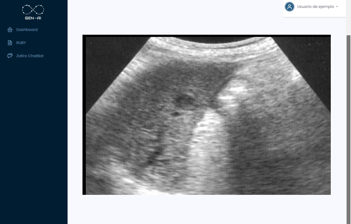
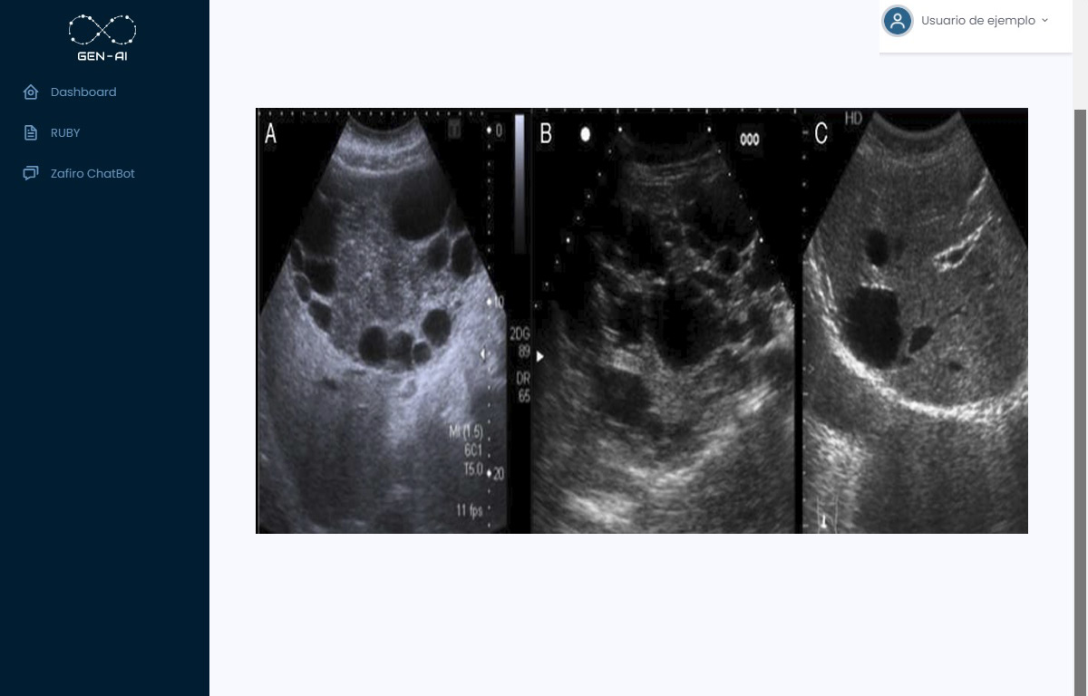
– In ultrasounds, it can report fatty liver, cysts, abnormalities in internal organs, etc.
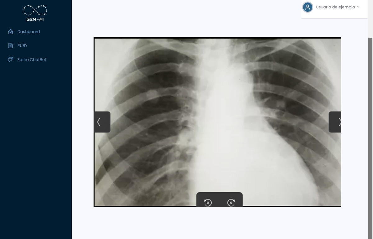
– In X-rays, it can identify opacities, tumors, fractures, signs of infection, among others.
– In CT scans or MRIs, it can detect brain lesions, vascular anomalies, tumor masses, etc.
- It generates a detailed report of the findings in each image.
- It provides recommendations based on the findings, such as additional studies, consultations with specialists, or lifestyle changes.
Output:
A report in JSON format detailing the findings image by image and the corresponding recommendations.
Example JSON Response:
{
   "diagnosis": {
      "image_1": "Hepatic steatosis (fatty liver) detected in the abdominal ultrasound.",
      "image_2": "Presence of an opacity in the left upper lobe of the lung, possible tumor mass.",
      "recommendations": [
         "A CT scan of the chest is suggested for a more detailed evaluation.",
         "Refer the patient to a pulmonologist.",
         "Advise the patient to quit smoking and adopt healthy lifestyle habits."
      ]
   }
}
Applications in Radiology and Medical Imaging
- Analysis of Chest Images:
– Description: The API can analyze chest X-rays to detect anomalies such as tumors, opacities, signs of infection, pneumonia, tuberculosis, among other pulmonary pathologies.
– Benefit: Helps radiologists identify potential pathologies more quickly and accurately, serving as a second opinion that complements their analysis.
- Diagnosis of Fatty Liver or Other Pathologies in Ultrasounds:
– Description: In abdominal ultrasounds, the API can identify signs of hepatic steatosis (fatty liver), cysts, gallstones, inflammation in organs, among other findings.
– Benefit: Provides support in the early diagnosis of liver diseases and other conditions, facilitating timely interventions.
- Identification of Fractures or Bone Damage in X-rays:
– Description: By analyzing bone X-rays, the API can detect fractures, fissures, osteoporosis, joint degeneration, and other musculoskeletal anomalies.
– Benefit: Improves the detection of injuries that might go unnoticed, especially in complex areas or in cases of microfractures.
- Detection of Brain Lesions in CT Scans and MRIs:
– Description: The API can analyze images from CT scans or MRIs to identify brain lesions, strokes, tumors, vascular anomalies, among others.
– Benefit: Aids in the rapid diagnosis of critical neurological conditions, where time is a determining factor.
Practical Examples of API Use
Example 1: Detection of Pneumonia in Chest X-rays
Request:
Instructions: “Analyze the chest X-rays for signs of pneumonia or pulmonary infections.”
Patient Data:
{
   "name": "María López",
   "age": "30",
   "gender": "Female",
   "medical_history": "Symptoms of fever and persistent cough for 5 days."
}
API Response:
{
   "diagnosis": {
      "image_1": "Interstitial infiltrates observed in both lungs, compatible with bilateral pneumonia.",
      "recommendations": [
         "Start antibiotic treatment as per protocol.",
         "Constant monitoring of vital signs and oxygen saturation.",
         "Follow-up with control X-rays in 48-72 hours."
      ]
   }
}
Example 2: Identification of Fractures in Bone X-rays
Request:
Instructions: “Analyze the X-rays of the right lower limb for fractures or bone injuries.”
Patient Data:
{
   "name": "Carlos Mendoza",
   "age": "25",
   "gender": "Male",
   "medical_history": "Recent sports injury, intense pain in the right leg."
}
API Response:
{
   "diagnosis": {
      "image_1": "An oblique fracture in the diaphysis of the right tibia is identified.",
      "recommendations": [
         "Immobilize the affected limb.",
         "Refer to orthopedics for surgical evaluation.",
         "Administer analgesics as needed."
      ]
   }
}
Example 3: Detection of Fatty Liver in Ultrasounds
Request:
Instructions: “Analyze the abdominal ultrasounds for liver abnormalities.”
Patient Data:
{
   "name": "Ana Torres",
   "age": "50",
   "gender": "Female",
   "medical_history": "Obesity, history of high cholesterol."
}
API Response:
{
   "diagnosis": {
      "image_1": "Liver echogenicity is increased, indicative of hepatic steatosis (fatty liver).",
      "recommendations": [
         "Recommend a low-fat diet and regular exercise.",
         "Monitor cholesterol and triglyceride levels.",
         "Follow-up with periodic ultrasounds to monitor changes."
      ]
   }
}
Advantages of Using the API in Radiology and Medical Imaging
- Support in Early Diagnosis:
– The API helps identify anomalies that could go unnoticed, allowing for more timely interventions and improving patient outcomes.
- Efficiency and Time Savings:
– Automates part of the analysis process, reducing the workload of radiologists and speeding up response times.
- Automated Second Opinion:
– Provides a second opinion that can complement the healthcare professional’s diagnosis, increasing accuracy and reliability.
- Personalization of Recommendations:
– Generates recommendations tailored to the patient, considering their medical history and clinical context.
- Does Not Store Sensitive Information:
– Ensures privacy and confidentiality, as it does not store images or personal data, only the generated report.
- Improvement in Patient Care:
– By streamlining the diagnosis process, treatment can be initiated more quickly, enhancing patient experience and satisfaction.
Summary
The API /virtualbot/analisys_image_report/ is an innovative tool for the Radiology and Medical Imaging sector, providing significant support to healthcare professionals in the analysis of medical images. By combining image information with the patient’s clinical history, the API generates detailed reports and personalized recommendations, facilitating more accurate and timely diagnoses. Its implementation in clinics and hospitals can enhance efficiency, reduce specialists’ workload, and, most importantly, contribute to better care and outcomes for patients.


